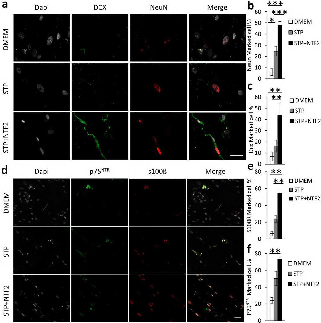
Fig. 6. Differentiation of hDPSCs grown in DMEM+10% FBS, STP and STP/NTF2 media to neuronal and glial-lineage cells: a) After a preconditioning growth phase of hDSPSCs in different culture media, neuronal and glial differentiation of hDPSCs was induced by culturing cells with the neural-inductive medium Neurocult Differentiation for 3 weeks. Cells were stained for the immature neuronal marker DCX (green), and mature neuronal marker NeuN (Red). Scale Bar 50µm. b-c) Graph showing positively marked cell percentage for NeuN and DCX. d) hDPSCs were stained with the Schwann cell markers, P75NTR (green) and S100ß (Red). Scale Bar 50µm. e-f) Graph showing percentage of positive cells for S100ß and P75NTR. All statistics were analyzed by ANOVA following by Scheffe post-hoc analysis *=p<0.05, **=p <0.01, ***p<0.001. Experiments were performed in triplicate.